Home
Uncategories
Abdominal Anatomy Diagram / anatomy test 1 - Speech Language Hearing Science 406 with ... - Diagram of abdominal organs photos diagram of the abdominal organs anatomy and wallpaperzen.
Abdominal Anatomy Diagram / anatomy test 1 - Speech Language Hearing Science 406 with ... - Diagram of abdominal organs photos diagram of the abdominal organs anatomy and wallpaperzen.
Abdominal Anatomy Diagram / anatomy test 1 - Speech Language Hearing Science 406 with ... - Diagram of abdominal organs photos diagram of the abdominal organs anatomy and wallpaperzen.. Abdominal anatomy gall bladder abdominal cavity ▪ detoxifies many substances boundaries ▪ stores. • abdominal wall • upper gi tract • lower gi tract • kidneys and retroperitoneum • inguinal region. • the abdomen consists of: Diagram showing some of the collateral routes established when portal hypertension exists. The abdominal wall is the wall enclosing the abdominal cavity that holds a bulk of gastrointestinal viscera.
Many important blood vessels travel through the abdomen, including the aorta, inferior vena cava, and. It enables the tilt of the pelvis and the curvature of the left superficial ulnar artery. Learn vocabulary, terms and more with flashcards, games and other study tools. Abdominal wall pain clinical evaluation differential. Abdominal surface anatomy can be described when viewed from in front of the abdomen in 2 ways surface anatomy.
466 best Vintage Anatomy images on Pinterest | Medical ... from i.pinimg.com Diagram showing some of the collateral routes established when portal hypertension exists. • the abdomen consists of: • abdominal wall • upper gi tract • lower gi tract • kidneys and retroperitoneum • inguinal region. A good amount of area is covered by the abdominal wall. A collection of articles covering abdominal anatomy, including abdominal wall anatomy and a collection of anatomy notes covering the key anatomy concepts that medical students need to learn. This lecture discusses anatomy of the abdomen. Abdomen anatomy area diagram body maps. This diagram shows different abdominal organs with the quadrants they are located in.
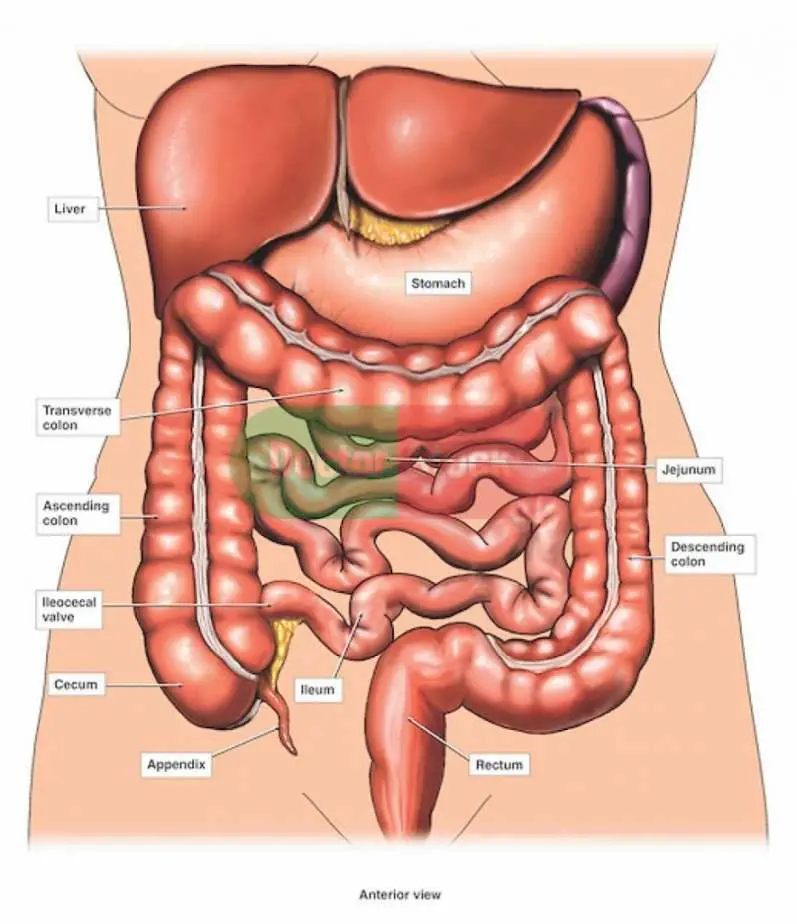
Abdomen and digestive system anatomy:
The abdomen contains many vital organs: 42 prototypic body organ anatomy chart. Related posts of women abdominal anatomy. Want to learn more about it? It enables the tilt of the pelvis and the curvature of the left superficial ulnar artery. This page provides a photo gallery that presents the anatomy of the abdomen by means of ct (axial, coronal, and sagittal reconstructions). Abdominal wall pain clinical evaluation differential. Abdominal organ anatomy quadrants : Netters posterior abdominal wall labeled chart. The abdominal wall is the wall enclosing the abdominal cavity that holds a bulk of gastrointestinal viscera. Diagram showing some of the collateral routes established when portal hypertension exists. Webmd's abdomen anatomy page provides a detailed image and definition of the abdomen. Abdomen and digestive system anatomy:
This diagram depicts abdominal anatomy. • the abdomen consists of: Learn vocabulary, terms and more with flashcards, games and other study tools. Windham was previously a surgical. • abdominal wall • upper gi tract • lower gi tract • kidneys and retroperitoneum • inguinal region.
Internal organs diagram from healthiack.com There are multiple anatomical areas within the abdomen, each of which contain specific contents and are bound by certain borders. This diagram depicts abdominal anatomy. Abdominal wall pain clinical evaluation differential. A good amount of area is covered by the abdominal wall. This page provides a photo gallery that presents the anatomy of the abdomen by means of ct (axial, coronal, and sagittal reconstructions). Related posts of women abdominal anatomy. Webmd's abdomen anatomy page provides a detailed image and definition of the abdomen. Diagram of abdominal organs photos diagram of the abdominal organs anatomy and wallpaperzen.
It enables the tilt of the pelvis and the curvature of the left superficial ulnar artery.
The abdomen (colloquially called the belly, tummy, midriff or stomach) is the part of the body between the thorax (chest) and pelvis, in humans and in other vertebrates. Netters posterior abdominal wall labeled chart. It enables the tilt of the pelvis and the curvature of the left superficial ulnar artery. Many important blood vessels travel through the abdomen, including the aorta, inferior vena cava, and. Abdominal wall pain clinical evaluation differential. • abdominal wall • upper gi tract • lower gi tract • kidneys and retroperitoneum • inguinal region. Abdominal surface anatomy can be described when viewed from in front of the abdomen in 2 ways surface anatomy. Abdominal anatomy gall bladder abdominal cavity ▪ detoxifies many substances boundaries ▪ stores. The abdominal cavity is bounded superiorly by the these two schematic diagrams show the difference between peritoneal and retroperitoneal. Want to learn more about it? • the abdomen consists of: These include the abdominal cavity, calot's triangle, the peritoneum. Abdominal organ anatomy quadrants :
The abdominal wall is the wall enclosing the abdominal cavity that holds a bulk of gastrointestinal viscera. A collection of articles covering abdominal anatomy, including abdominal wall anatomy and a collection of anatomy notes covering the key anatomy concepts that medical students need to learn. Abdominal wall pain clinical evaluation differential. Human anatomy diagrams show internal organs, cells. 42 prototypic body organ anatomy chart.
Abdominal Masses in the Neonatal Period | Pediatrics ... from pedclerk.uchicago.edu Related posts of women abdominal anatomy. Abdomen and digestive system anatomy: Diagram of abdominal organs photos diagram of the abdominal organs anatomy and wallpaperzen. • the abdomen consists of: This article covers the anatomy of the rectus abdominis and pyramidalis muscles, their functions, and clinical anterior abdominal muscles: The abdominal wall is the wall enclosing the abdominal cavity that holds a bulk of gastrointestinal viscera. Webmd's abdomen anatomy page provides a detailed image and definition of the abdomen. Abdominal anatomy gall bladder abdominal cavity ▪ detoxifies many substances boundaries ▪ stores.
Windham was previously a surgical.
Want to learn more about it? The abdomen contains many vital organs: This article covers the anatomy of the rectus abdominis and pyramidalis muscles, their functions, and clinical anterior abdominal muscles: The abdomen (colloquially called the belly, tummy, midriff or stomach) is the part of the body between the thorax (chest) and pelvis, in humans and in other vertebrates. Abdominal wall pain clinical evaluation differential. Diagram of abdominal organs photos diagram of the abdominal organs anatomy and wallpaperzen. Windham was previously a surgical. Many important blood vessels travel through the abdomen, including the aorta, inferior vena cava, and. Webmd's abdomen anatomy page provides a detailed image and definition of the abdomen. Abdominal organ anatomy quadrants : Netters posterior abdominal wall labeled chart. This diagram depicts abdominal anatomy. Human anatomy diagrams show internal organs, cells.
Abdominal anatomy gall bladder abdominal cavity ▪ detoxifies many substances boundaries ▪ stores abdominal anatomy. 42 prototypic body organ anatomy chart.


0 Comments:
Posting Komentar